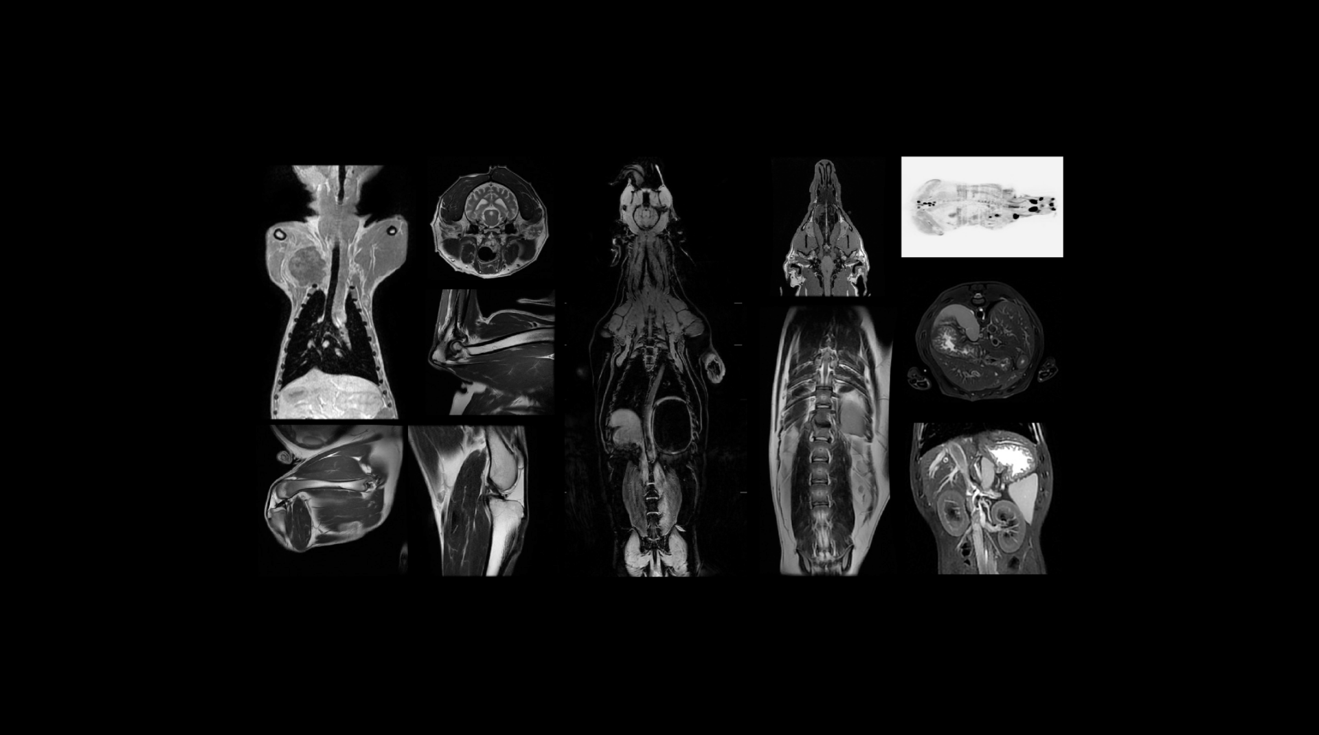
MRI Image Quality Improvement

Sharper images, Faster scans, Better care.
MRI Quality

MRI image quality improvement

High-quality MRI is essential for accurate diagnosis and reliable research in veterinary and preclinical settings. Precise imaging enables veterinarians to detect subtle abnormalities, guide treatment plans, and monitor disease progression. In research, high-quality images ensure precise biomarker analysis, improving the reliability of data for drug and device development.

However, veterinary and preclinical MRI presents unique challenges. The smaller anatomical structures of animals require higher resolution to capture fine details, but this can increase scan times, raising the risk of anesthesia complications. Additionally, patient movement—whether due to respiration, stress, or anesthesia limitations—can introduce artifacts that degrade image clarity.

These challenges necessitate specialized tools and workflows, such as AI-powered post-processing, optimized imaging protocols, and efficient positioning systems.

The role of MRI engineers and radiologists in enhancing image quality

Optimizing MRI image quality demands close collaboration between MRI engineers and radiologists. Both play critical roles in fine-tuning imaging parameters, refining protocols, and ensuring consistent performance across various MRI systems.

Together, they address challenges like balancing image resolution with scan speed and maintaining clear contrast between tissues, which is especially important in neurology and oncology.

For instance, adjusting slice thickness can improve anatomical detail for smaller animals, but this may also increase scan time. Radiologists and engineers must carefully balance these factors to minimize patient stress and anesthesia use.

Other critical adjustments include ensuring optimal contrast settings to distinguish between soft tissues and managing the Signal-to-Noise Ratio (SNR) to maintain image clarity.

Showing images

Optimizing parameters for precision

Resolution: Higher resolution is essential for smaller animals to capture minute details, though it increases scan time. Balancing resolution with speed is crucial to avoid prolonged anesthesia exposure.

Contrast and Signal-to-Noise Ratio (SNR): Contrast adjustments help differentiate soft tissues or lesions, while SNR ensures image clarity by reducing background noise.

Slice Thickness and Orientation: Thinner slices provide more anatomical detail but can lead to longer acquisition times and more artifacts. Engineers and radiologists collaborate to find the right balance, depending on the clinical or research focus.

Collaboration to refine protocols
Tailoring MRI protocols to specific research or diagnostic needs ensures that image quality remains high across different applications, such as neurological imaging or musculoskeletal assessments.

Engineers handle the technical aspects, including calibrating machines and configuring coils, while radiologists determine anatomical targets and clinical priorities.

Standardized tools like HawkProtocols offer a streamlined framework, helping facilities maintain consistency and efficiency.

Case Study: Addressing motion artifacts with optimized protocols
Motion artifacts are a common challenge in veterinary MRI, often caused by involuntary movements or respiration. Faster imaging techniques, such as Fast Spin Echo (FSE) or parallel imaging, help minimize scan times and reduce the need for extended anesthesia. 

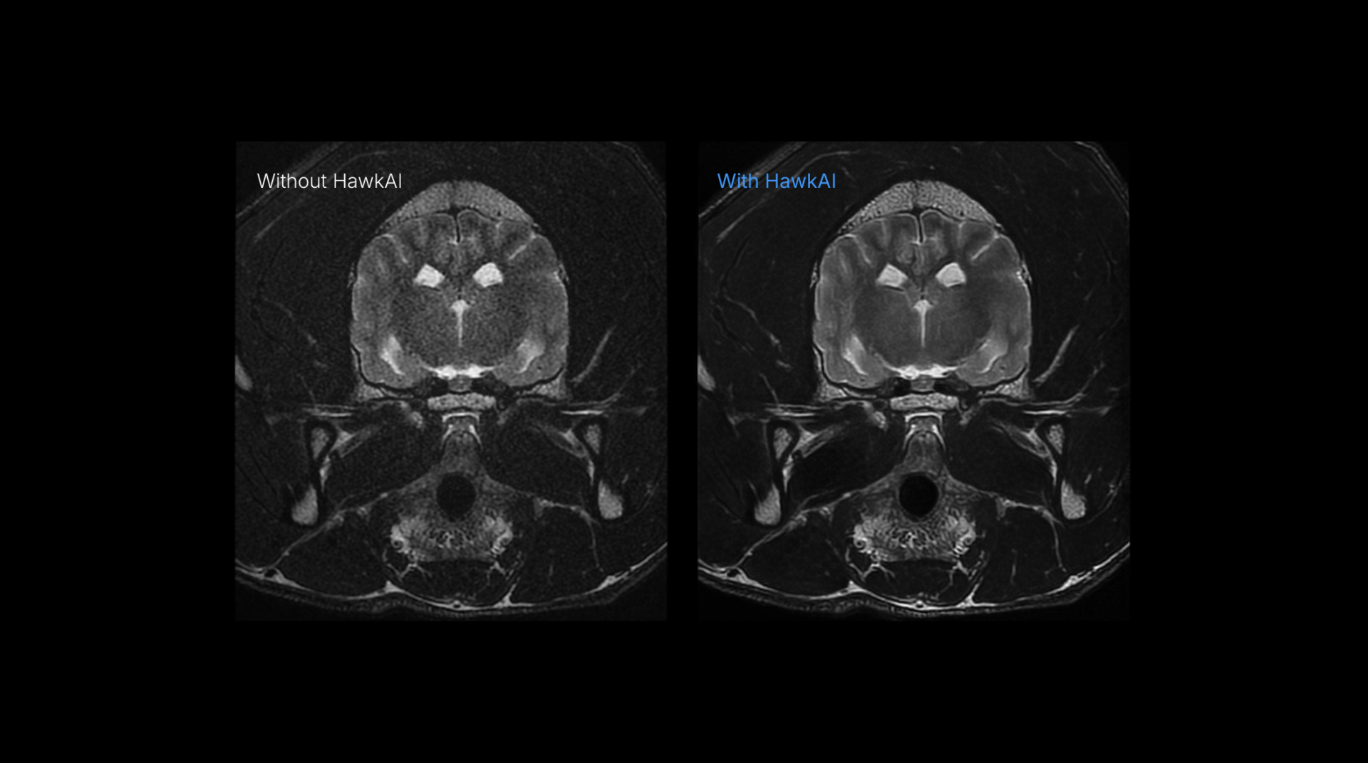
In addition, AI-based tools like HawkAI can correct noise and artifacts during post-processing, enhancing image clarity even when faster acquisition compromises resolution.

This integrated approach ensures veterinarians and researchers capture the highest quality images while minimizing patient risk.

Since we have our own MRI, we have a clinical component and can participate in preclinical studies. These activities allow our application engineers to remain relevant and aligned with practices when optimizing protocols.

Incorporating advanced technologies to overcome MRI limitations

Modern technologies help address the limitations of older MRI systems, reduce artifacts, and improve imaging precision. By integrating AI-powered tools and optimized workflows, clinics and researchers can achieve better diagnostic accuracy while enhancing efficiency and patient safety.

AI-Driven Post-Processing with HawkAI

HawkAI enhances MRI performance by automating noise reduction, improving SNR, and boosting resolution. This is also beneficial for older MRI systems, as it elevates their performance to match that of newer models.

Facilities can achieve up to 30% improvement in image clarity, reducing the need for hardware upgrades. Additionally, shorter scan times—enabled by HawkAI’s optimized algorithms—reduce anesthesia exposure, improving both patient safety and workflow efficiency.

Improving patient positioning with HawkSpine

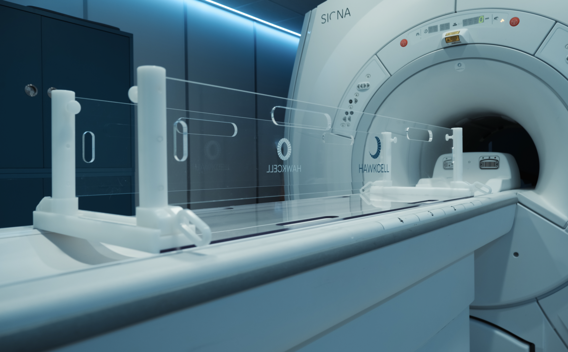
Correct positioning plays a significant role in preventing motion artifacts and ensuring consistent image quality. HawSpine offers an adjustable and ergonomic solution for positioning animals of varying sizes. 

It simplifies setup, reducing preparation time to under two minutes, and minimizes stress for both the animal and the technician. This streamlined process decreases the chance of motion-related artifacts and reduces the need for repeat scans.

HawkSpine

Standardization with HawkProtocols

Pre-designed protocols from HawkProtocols ensure uniform image quality across different MRI systems and operators. These expert-curated protocols cover various applications—ranging from brain to orthopedic imaging—ensuring that facilities maintain diagnostic accuracy and research consistency.

Standardized protocols not only improve efficiency but also reduce the likelihood of operator error.

HawkProtocols

The long-term benefits of improved MRI image quality

Clinical Impact

High-quality imaging allows veterinarians to make timely and accurate diagnoses, reducing unnecessary procedures and treatment delays. This helps lower healthcare costs and improves patient outcomes by enabling targeted interventions.

Research Benefits

In preclinical research, clear MRI images are essential for precise biomarker identification and reliable data collection. High-quality imaging improves the reproducibility of research results, which is critical for drug development and regulatory approval.

Faster, more accurate imaging also accelerates the pace of scientific innovation, helping researchers bring new treatments to market more efficiently.

Ethical Impact

Improving image quality directly supports animal welfare by reducing the need for repeat scans and minimizing anesthesia exposure. Shorter scan times also lessen the stress experienced by animals, aligning MRI practices with ethical standards and best practices for care.

High-quality imaging thus not only benefits research and diagnostics but also fosters the humane treatment of animal patients.

Conclusion

Achieving excellence in MRI imaging requires a combination of optimized parameters, advanced technologies, and collaboration among veterinarians, radiologists, and MRI engineers. Tools like HawkAI, HawkSpine, and HawkProtocols empower professionals to improve diagnostic precision and research outcomes while enhancing workflow efficiency.

By investing in high-quality imaging practices and leveraging cutting-edge technologies, clinics, and research facilities can push the boundaries of veterinary and preclinical MRI. HawkCell is committed to supporting this pursuit by providing innovative tools and expertise tailored to meet the evolving demands of these fields.

Try us now

Schedule a discovery call with one of our representatives

    HawkCell