Precision in preclinical MRI

We deliver efficacy and safety studies at the same time.
Bio / Med Tech

Accelerate drug development with HawkCell’s advanced MRI biomarkers and preclinical solutions

HawkCell offers innovative solutions for Biotech and Medtech companies, specializing in MRI biomarkers and preclinical MRI studies. Our comprehensive approach enables you to conduct efficacy and safety studies simultaneously, accelerating your drug development process.

Key benefits

Efficiency

Conduct multiple studies in parallel, saving time and resources.

Precision

Utilize MRI biomarkers for accurate measurement of disease progression and treatment response.

Safety

Assess the safety profile of your drug candidates early in development.

Data-driven decisions

Gain valuable insights from our preclinical MRI studies to inform your drug development strategy.

Revolutionizing research with MRI

Faster

MRI significantly speeds up preclinical research by offering real-time, non-invasive, high-resolution imaging of biological processes. This enables researchers to:

Monitor disease progression and treatment response.

Evaluate drug efficacy in targeting specific biological pathways.

Study anatomical changes in tissue structure and function.

Reduce animal use by minimizing the number of animals required through comprehensive data from a single scan.

Access in-vivo information

MRI provides detailed, real-time imaging of biological processes within living organisms, allowing researchers to study organ, tissue, and cellular structure and function without invasive procedures. This yields valuable insights into disease mechanisms and drug responses.

Better ROI

MRI data helps identify safety concerns or inefficacy early, reducing costly clinical trial failures. Additionally, MRI-derived data strengthens patent applications, enhancing protection of intellectual property.

More ethical

MRI’s non-invasive nature reduces the need for multiple animals in research, promoting more ethical studies by minimizing animal suffering and eliminating the pain associated with invasive techniques.

Our services

MRI biomarker development

We develop MRI biomarkers tailored to answer specific therapeutic areas and research needs.

Preclinical MRI studies

Our experienced team conducts comprehensive preclinical MRI studies, including efficacy, safety, and pharmacokinetic assessments.

Data analysis and interpretation

We provide expert analysis and interpretation of MRI data, delivering actionable insights.

Our MRI biomarkers ⬇️

Whole Body Diffusion Imaging (WBDI)

Whole Body Diffusion Imaging (WBDI) is a specialized MRI technique that provides detailed information about tissue microstructure and cellular organization throughout the entire body. By measuring the diffusion of water molecules within tissues, WBDI can detect abnormalities in cellular architecture, such as those associated with diseases like cancer and metastases.

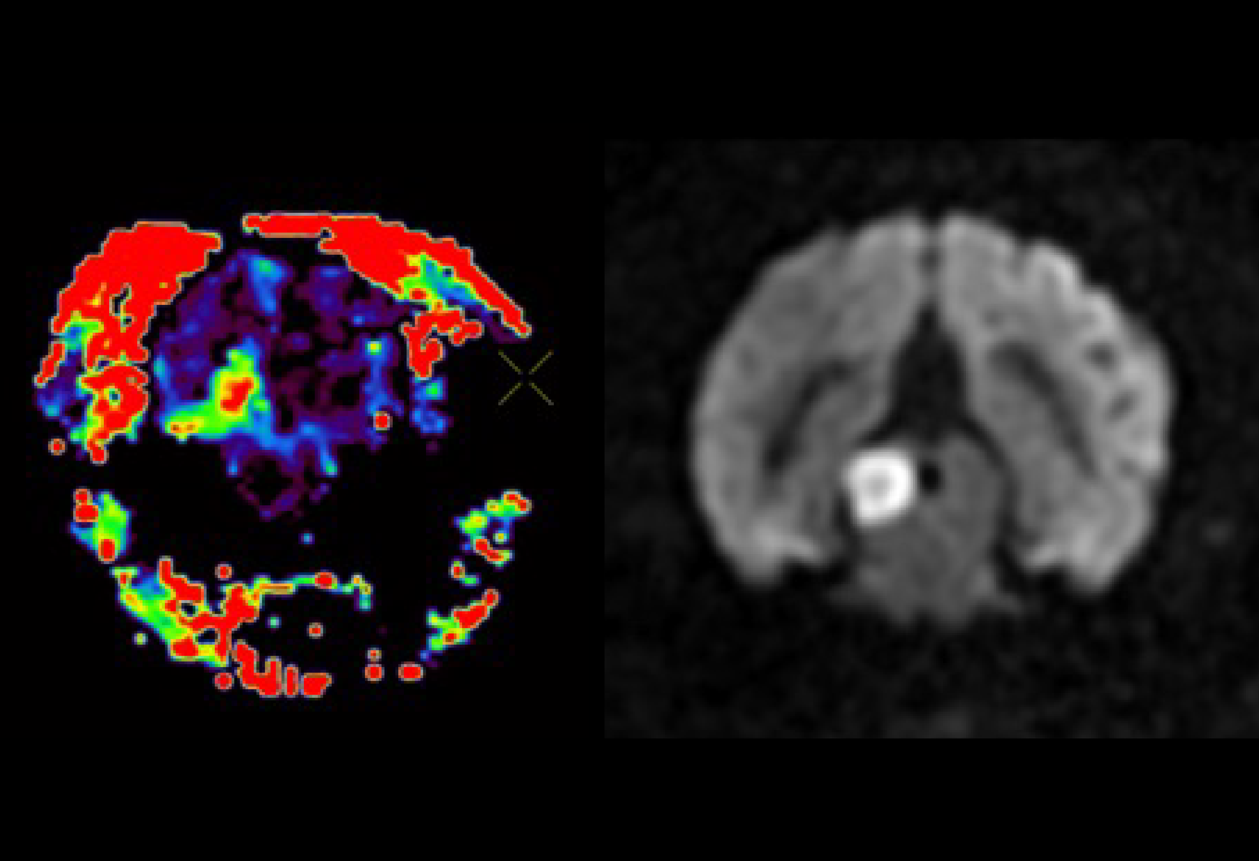
Perfusion MRI

Perfusion MRI measures blood flow within tissues. By quantifying the rate at which blood enters and exits a particular region, perfusion MRI offers valuable insights into tissue metabolism, oxygen delivery, and vascular function, particularly useful for assessing conditions like ischemic stroke.

Perfusion MRI

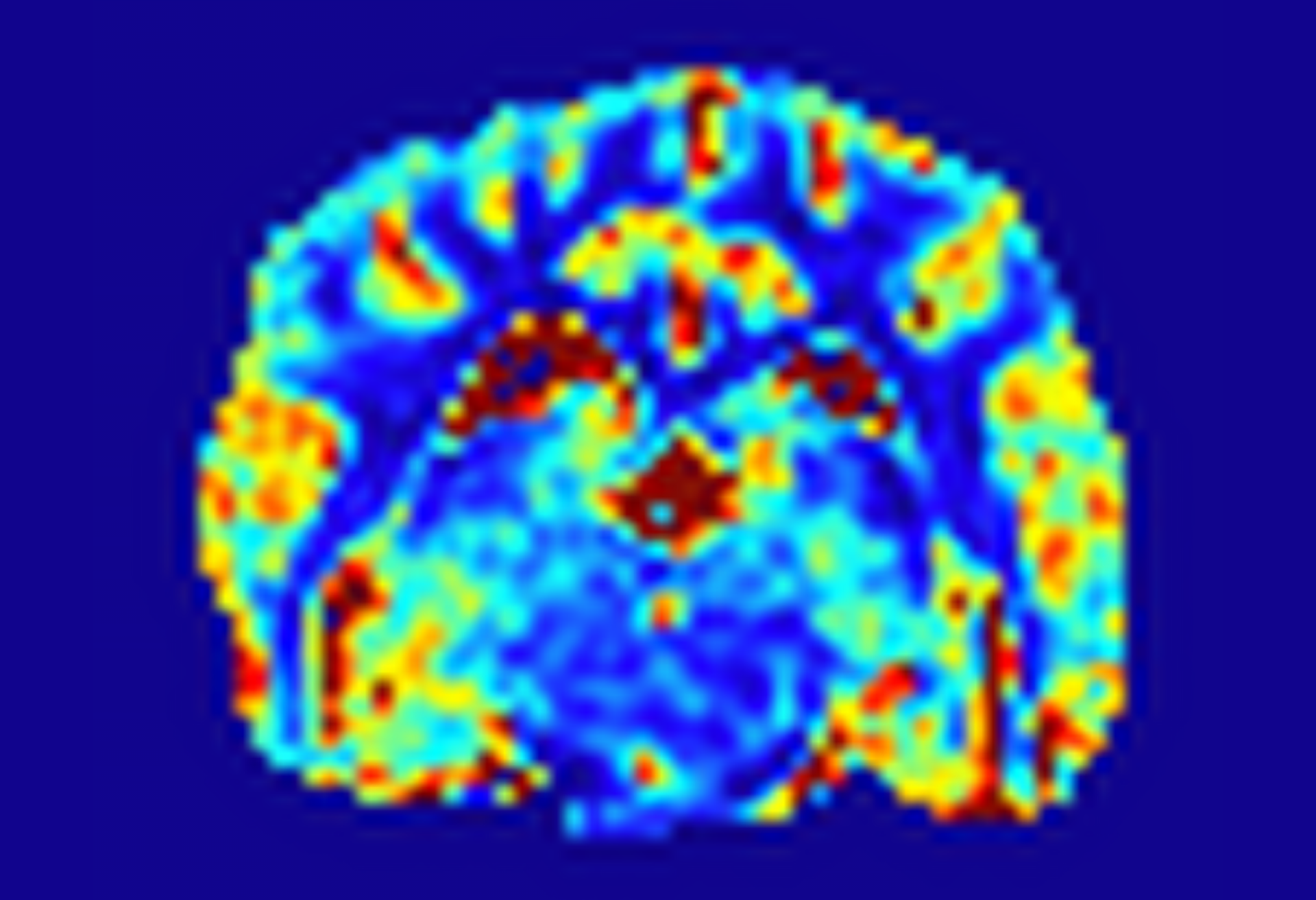
T1 Mapping

T1 Mapping is an MRI technique that measures the longitudinal relaxation time, indicating the time it takes for the magnetization of protons to recover to 63% of its equilibrium after a radiofrequency pulse. T1-weighted images are often used to visualize anatomical structures and differentiate between tissues with varying water content.

T1

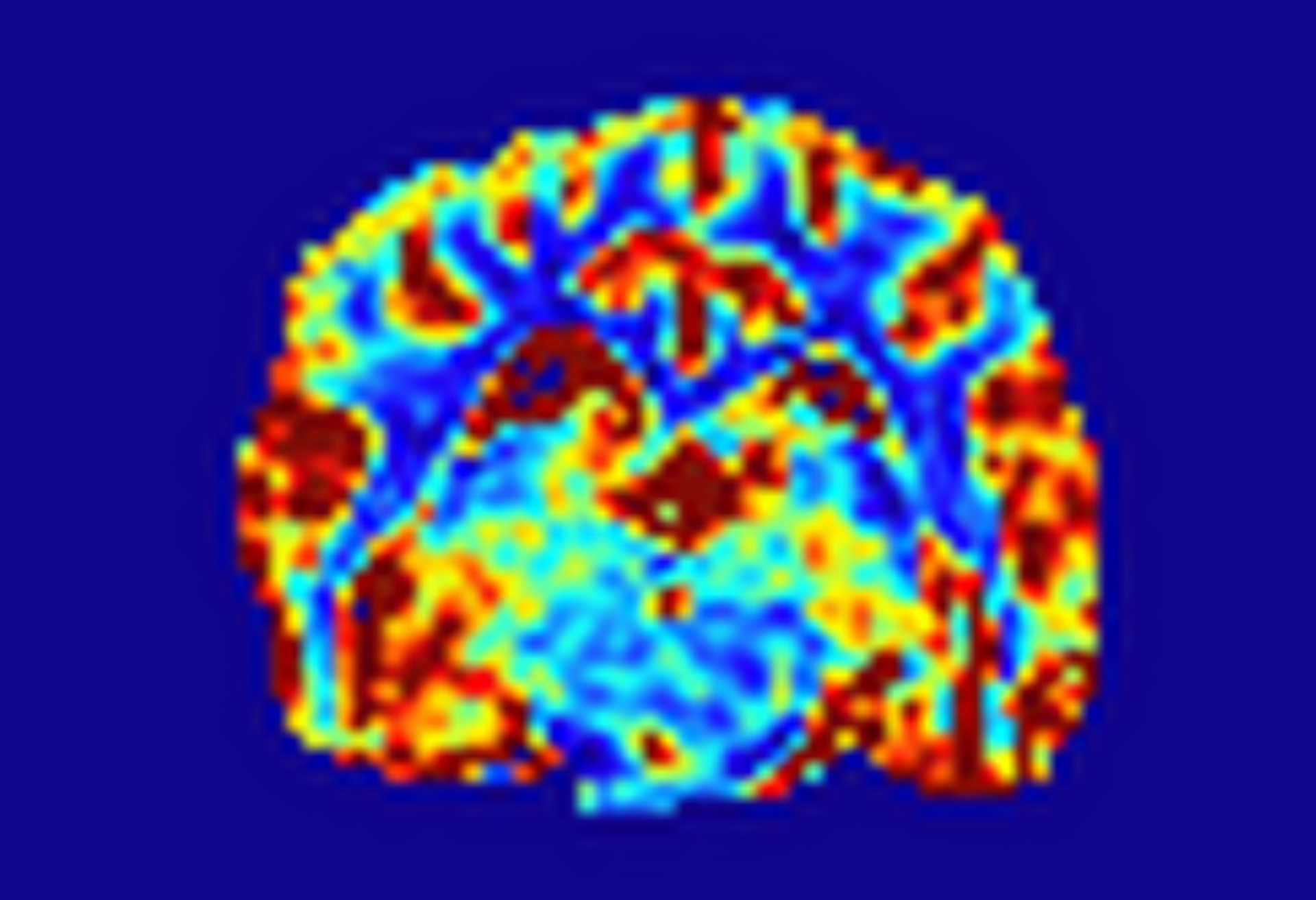
T2 Mapping

T2 Mapping measures the transverse relaxation time, which is the time it takes for proton magnetization to decay to 37% of its initial value after a radiofrequency pulse. T2-weighted images are commonly used to visualize fluid-filled structures and detect abnormalities in tissue microstructure.


T2

R2 Mapping*

R2 Mapping* measures the rate of transverse relaxation of tissue protons, influenced by factors like magnetic field inhomogeneities, susceptibility effects, and iron content in tissues.

Quantitative Susceptibility Mapping (QSM)

Quantitative Susceptibility Mapping (QSM) uses advanced post-processing algorithms to reconstruct quantitative susceptibility maps from raw phase images, providing precise estimates of magnetic susceptibility within each voxel. Unlike R2* mapping, QSM differentiates between paramagnetic and diamagnetic inclusions, making it particularly useful in neurodegenerative diseases where changes in tissue myelin content (diamagnetic) and iron content (paramagnetic) may coexist.

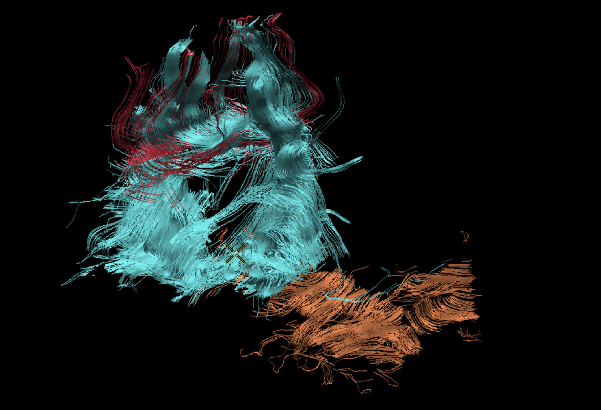
Diffusion Tensor Imaging (DTI)

Diffusion Tensor Imaging (DTI) estimates the organization of axonal (white matter) structures through anisotropic diffusion. Using fiber tracking techniques, DTI data allows for the 3D reconstruction of neural tracts, providing valuable insights into brain connectivity.

Our development pipeline

For preclinical and clinical quantitative MRI imaging biomarkers.
Neurology, oncology, rhumatology, urology, gastrointestinal

For more information about our development pipeline for preclinical and clinical quantitative MRI imaging biomarkers, please send us a request.

Our other services

For Academia

Enhance image quality and reduce scan time.

Learn more
HawkAI

For CRO

Experience seamless preclinical MRI research.

Learn more
Mobile MRI

Try us now

Schedule a discovery call with one of our representatives

    HawkCell