Abstract
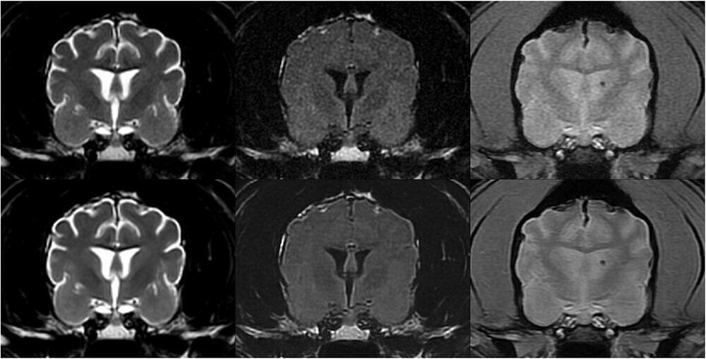
In this analytical cross-sectional method comparison study, we evaluated brain MR images in 30 dogs and cats with and without using a DICOM-based deep-learning (DL) denoising algorithm developed specifically for veterinary patients. Quantitative comparison was performed by measuring signal-to-noise (SNR) and contrast-to-noise ratios (CNR) on the same T2-weighted (T2W), T2-FLAIR, and Gradient Echo (GRE) MR brain images in each patient (native images and after denoising) in identical regions of interest.
Qualitative comparisons were then conducted: three experienced veterinary radiologists independently evaluated each patient’s T2W, T2-FLAIR, and GRE image series. Native and denoised images were evaluated separately, with observers blinded to the type of images they were assessing. For each image type (native and denoised) and pulse sequence type image, they assigned a subjective grade of coarseness, contrast, and overall quality.
For all image series tested (T2W, T2-FLAIR, and GRE), the SNRs of cortical gray matter, subcortical white matter, deep gray matter, and internal capsule were statistically significantly higher on images treated with DL denoising algorithm than native images. Similarly, for all image series types tested, the CNRs between cortical gray and white matter and between deep gray matter and internal capsule were significantly higher on DL algorithm-treated images than native images.
The qualitative analysis confirmed these results, with generally better coarseness, contrast, and overall quality scores for the images treated with the DL denoising algorithm.
In this study, this DICOM-based DL denoising algorithm reduced noise in 1.5T MRI canine and feline brain images, and radiologists’ perceived image quality improved.