Introduction
Preclinical models, such as murine models, are essential for evaluating the efficacy of therapeutic treatment in oncology research. Recently, novel murine models with surgically implanted orthotopic tumor allow a more accurate representation of human cancer than traditional subcutaneous models. Orthotopic models however require in vivo evaluation. This shift necessitates the use of non-invasive imaging techniques to monitor tumor growth, as caliper measurements used for subcutaneous tumors are no longer applicable.
Monitoring the tumoral volume or its follow-up is often imprecise. It is also difficult to attribute weight changes to tumor progression or other confounding factors. One of the key endpoints in these studies is the assessment of tumoral volume, which allows researchers to monitor not only tumor growth but also response to treatments over time. MRI has emerged as a non-invasive and precise imaging technique providing detailed anatomical and functional information, making it particularly useful in oncology research.
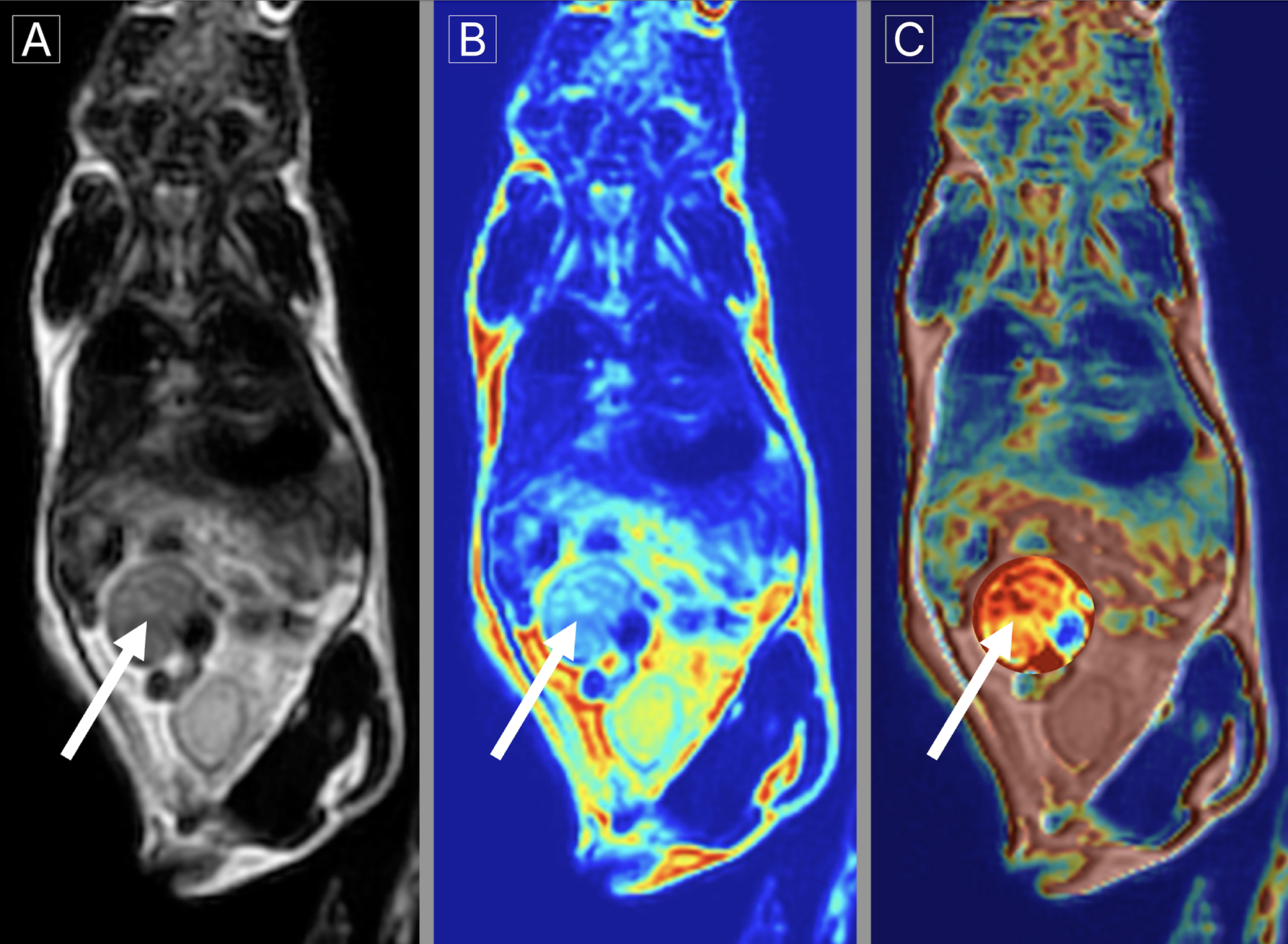
This white paper, meant as a proof of concept, discusses the pertinence of using MRI technology to assess tumoral volume in an orthotopic murine colorectal cancer model.


